Medical Tech Outlook Weekly Brief
Be first to read the latest tech news, Industry Leader's Insights, and CIO interviews of medium and large enterprises exclusively from Medical Tech Outlook
A featured contribution from Leadership Perspectives: a curated forum reserved for leaders nominated by our subscribers and vetted by our MedTech Outlook Advisory Board.


Technologic advancements in orthopaedic and spine surgery have evolved in a variety of directions, and they are providing for better care than ever for our patients. One of the fascinating and increasingly useful new tools has been adopted from other industries---3-D printing. This particular technology has allowed engineers and clinicians to develop implants that are more powerful and specific for their application. Imagine a patient who has a tumor that enveloped a portion of their skeleton, and treatment requires a resection of that critical part of their structure. Or, consider a patient who has lost a portion of their skull or face to an unfortunate traumatic event. These circumstances have historically been challenging to manage for a variety of reasons. Being able to create a structurally-sound and biologically-friendly reconstructive element to replace this lost anatomy was technically challenging, and the timeliness of both producing it to specification, as well as overcoming the regulatory burdens in doing so were nearly insurmountable. Additionally, the cost of producing these individualized solutions generated a headwind that made them largely untenable.
Just as it has revolutionized other sectors, 3-D printing in the realm of skeletal reconstruction allows for modulation in the architecture and bioactivity of implants that was historically unavailable. Importantly, the lower production cost and patient customization of these implants thrusts us into a new frontier of more optimal and cost-efficient care provision. We now have the capacity to produce sizes and shapes of implants that can perfectly reconstitute lost skeletal anatomy, and the speed with which these devices can be prepared is achieving alignment with the clinical need.
Another important feature of this technology in making it attractive for skeletal reconstruction is the ability to modify not only the shape and size, but the architecture of the material. The biology of bone healing and harnessing its potential requires that implants mimic the features inherent to normal bone. Creating ideal pore size and structure on the surface, as well as internally, allows bone cells (osteoblasts, osteoclasts, and osteocytes) to facilitate a normal integration and healing process. We have known and used this concept for many decades to create ingrowth prostheses, such as with total hip replacements, however, generating the proper surface alone has not been sufficient in other areas, such as spine surgery. 3-D printing specifically provides the capacity to create the proper porosity and structural conditions internal to the implant, and thus more aptly provides a conductive effect for bone to grow through the implant. Ultimately, a more reliable integration can ensue.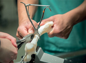
One of the great challenges in reconstructing skeletal elements that have been injured or diseased has been appropriately matching the mechanical properties required for function. Using nonbiologic materials, like metal or plastic, presents a problem in that they have different levels of hardness or stiffness than the bone they are replacing. This differential leads to frequent mechanical failures, including fractures or undesirable settling of the implant into the surrounding bone. Fortunately, the availability of computer algorithms and 3-D printing have been combined to solve this dilemma. We now have the capacity to modify the relative stiffness of structural implants by adjusting their architecture through these computer models. Thus, we can take a material, like titanium, which has a modulus of elasticity (stiffness) that is considerably greater than bone, and create an implant made of it with a relative stiffness that is equivalent to or even less than the surrounding bone. Conceptually, this reduction in stiffness would be somewhat like creating a spider-web with steel.
Nanotechnology has also found its way into osteobiologics. Through basic science research, it has been shown that cellular messaging and bone healing can be modulated through the topographical shapes and sizes of the surface of titanium, specifically (and potentially other materials). Harnessing this information has led to spinal implants, for instance, that can now encourage bone producing cells to communicate more effectively with one another in generating an improved bone healing and fusing environment following reconstruction.
Check Out:- MdTech Review
In combining these various technologic advancements, implants can be tailored to specific anatomic sizes and locations, with mechanical properties that can be further customized in accommodating differential bone strengths; and finally, with surface characteristics that further augment bone healing capacity. The resultant state-of-the-art implants advance patient care tremendously.